Tuesday 23 April 2024

Isolating and selecting microscope specimens

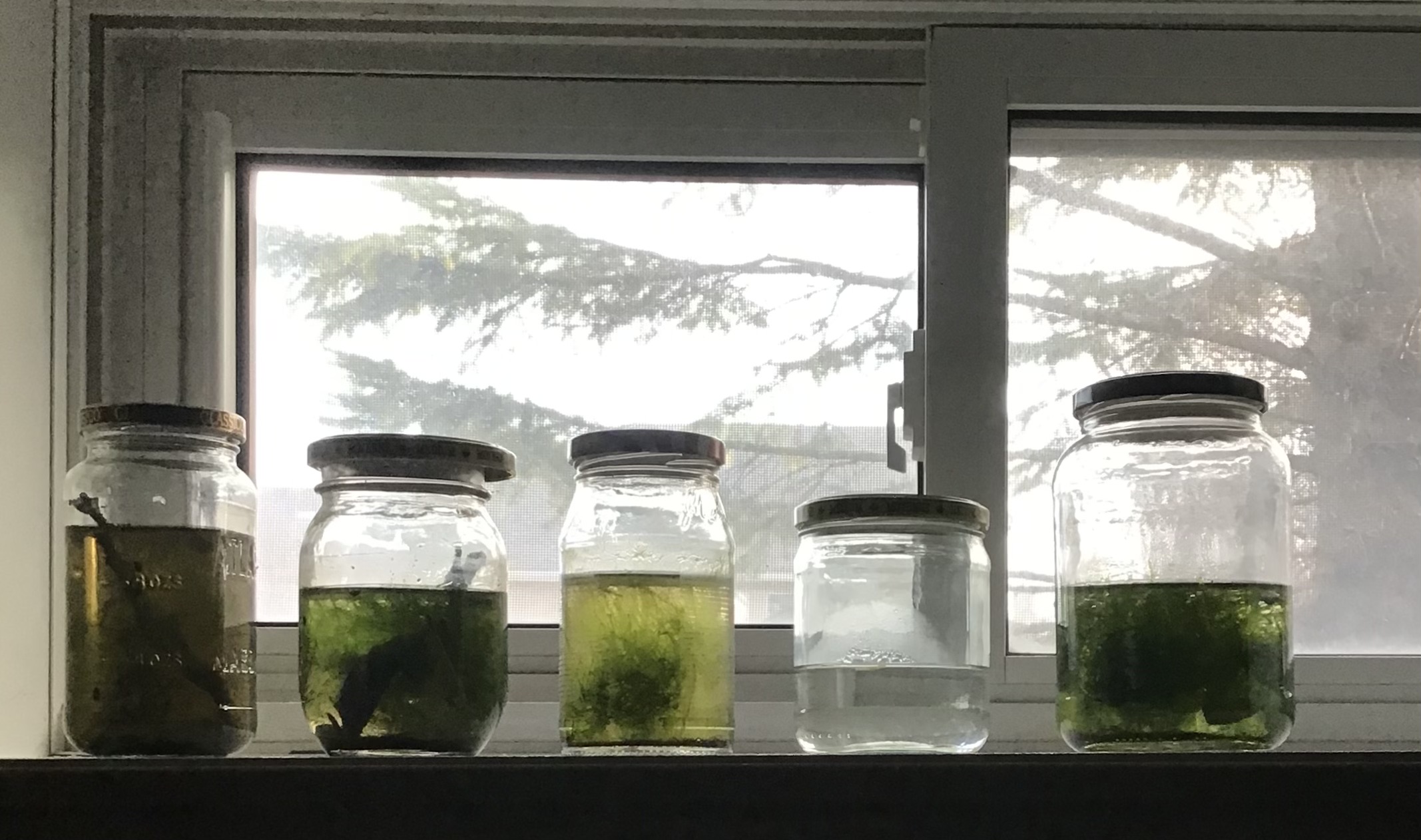
Deliberately selecting specimens to view under a microscope has always been a challenge for me...but I'm getting better! I thought I'd share my methodology in the hope that someone else can benefit from my experience. But before I get to the basics of what I do it's important to share that reading about techniques will only get you so far, you have to practice to get better. In my experience, the more you practice, the better you get.


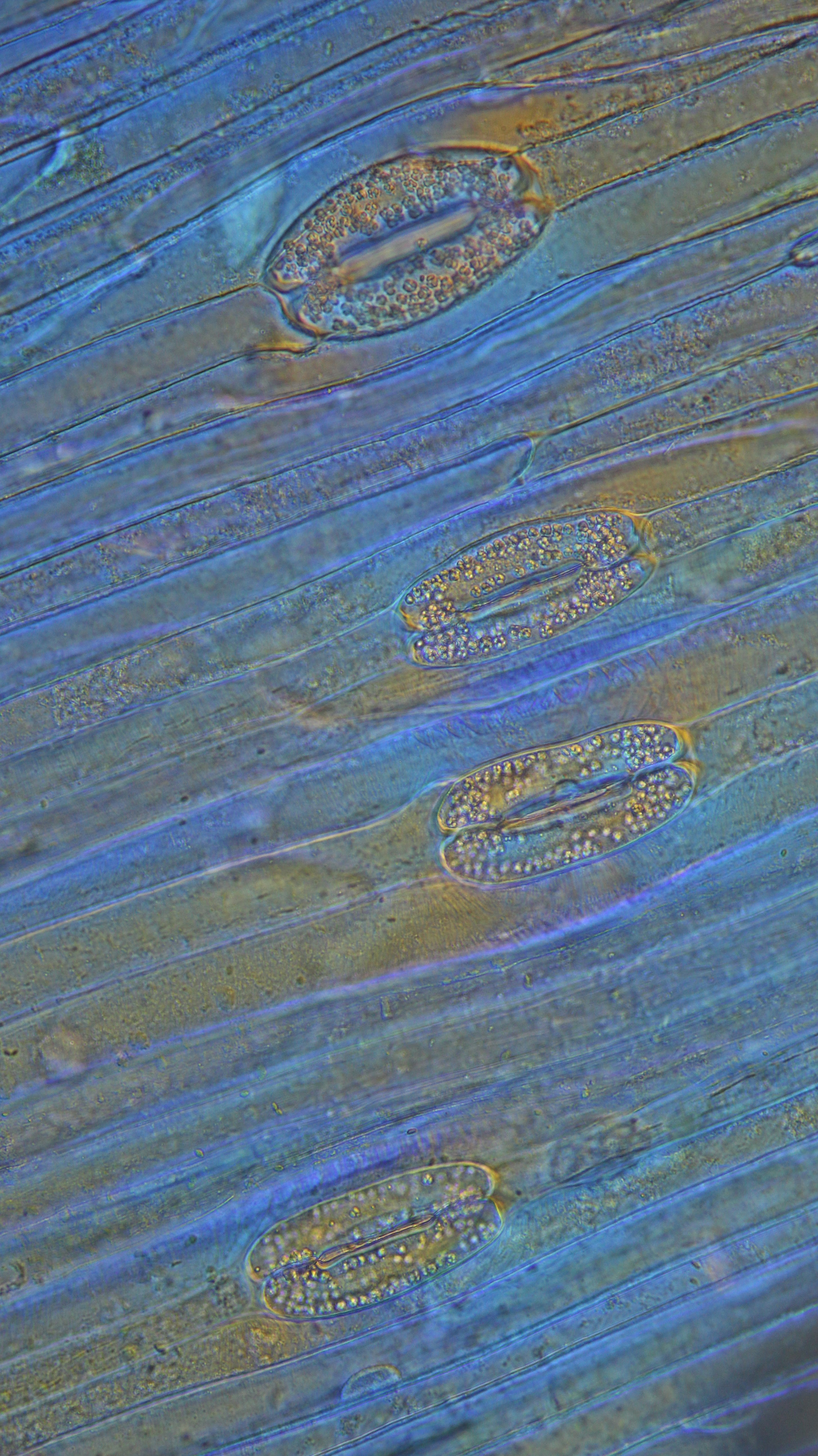
There are two methods I employ, one involves a stereoscope, the other a microscope. My favorite method employs the stereoscope, an LED goose neck lamp and a watch glass. I add 5 to 6 ml of water to my watch glass, apply some side lighting with the LED lamp and start hunting for specimens. My zoom stereoscope can employ powers ranging from 7X to 180X by using various accessory lenses but I generally work in the 14X to 90X range to isolate specimens. First, using a higher power I dial in to a critter that I want to examine with the microscope. possibly clean away debris with a needle, and suck up the specimen using a micro-pipette (20 to 40 ul) with a rubber bulb. This is then transferred to another area of the watch glass and viewed to confirm a successful transfer. If unsuccessful, repeat. When you have the specimen in a water drop, transfer it to a slide, put on a cover slip and start viewing under the microscope.


For those that don't own a stereoscope a similar technique can be employed using your microscope. In this case, a small volume of water, perhaps a couple of ml, is picked up with a dropper or micro-pipette and ejected along the length of a slide. Your specimen is then searched for under a low power, say 4X or 5X, and once found treated just like the critter in the previous paragraph.


Bonus tips: Cover slips float and specimens like to attach themselves to the bottom of them. I use the illustrated tweezers to drop them and pick them up.  


And stereoscopes can be as much fun to view protists as microscopes are. 3D and really fun to watch once you become familiar with what you're looking at.

Sunday 21 April 2024

Euglenids exibiting metaboly and flagellum motility

Euglena

  One of the jars I had been sampling from all winter           became quite green. I took a fairly large sample from the mid jar water and included some bottom sediment and ran it through the centrifuge at 800 RPM for about ten minutes. Out of the resulting pellet came a significant number of algae, a large number of which were various species of euglenids.


Phacus


There are now reports of over 1500 species so identification for the layperson is almost impossible.

But I did find some interesting movement types of one particularly numerous species, probably from Family Peranemidae, showing both metaboly and flagellum motility.


Also an image of a similar group in darkfield.


Monday 8 April 2024

Pass the Pollen, Honey


I watched a video about pollen extraction by Oliver of MicrobeHunter fame yesterday and thought it might be fun to duplicate what he did on his live-stream. He essentially put a small (2 ml) sample of honey with about 10 ml of water into a 15 ml vial and thoroughly mixed up the two into solution. Then about a 5 minute spin in a centrifuge at 4000 RPM, pour off the liquid and drop the pellet onto a slide with cover-slip. The result was a number of different pollen grains and at least one insect part, which according to Oliver, at one time belonged to a bee. All the pollen cells I found were in the range of 7 to 40 um long.


I also tried stacking a couple of the images with Picolay.


And here is the insect part, ostensibly from a bee. The piece is about 1/10 mm long.


Spring, old specimen jars and new opportunities.

After a long winter of limited access to fresh specimens, my river is again open, at least near the banks. However, the winter was an enlightening season since it taught me much about protist cultures, how to maintain them and how to sample them.


It all started last fall when I filled a standard “fish bowl” with water, mud and a few plants taken from the small river I live on. I began with a relatively large collection of microscopic specimens, including some larger types like gammarus, copepods, ostracods, fairy shrimp and daphnia. The bowl was placed in an east facing windowsill where it received direct morning light. This little micro environment provided a large number of critters for observation but after some time the plants died and things slowed down. Luckily I had a 30 gallon tank that had gone wild and in cleaning it up I was able to add a large matt of algae to my bowl, again rejuvenating it.

The fish bowl on the extreme left was the container that started it all

It was at that point I started a jar collection, seeding them with soil, water and algae from the fish bowl. These jars were “fed” with dried oak leaves, dead grasses from under the snow, vegetable scraps and various grains, cereals or even garden dirt. Most of these jars developed in different ways and provided a source of interesting, but limited, species over the winter. Another jar collection was started in a north facing window with none getting direct light. I had noticed the jars on the sunny sill were getting quite warm on clear days.

My north facing jars

One jar in which I was doing a Walstad experiment developed an algae explosion that I used to feed several of the jars with larger inhabitants. What I found interesting was that some of my best microbe sources were the smallest containers I  used, old 35 mm film containers. I even had a population explosion of several ciliate species under a cover slip on a slide I was keeping in a “wet” container to watch the development of snail eggs. 


After providing a winter of enjoyment under the microscope the jars will soon be emptied back into the river, cleaned up and readied for some new, springtime populations.

Sunday 31 March 2024

Video of a Rotifer

 I went back to the water sample from yesterday's post with the intent of getting better pictures to help with identifying this little critter. After several attempts I gave up and went the video route. I've tried going through several keys to ID her but I appear to have little talent in this area. Specimen is about 250 um long.

If you have any ideas please post them in the comments. Ive been told by an expert this specimen belongs to the Order Ploima, and possibly (slight) to the Genus Proales.


Saturday 30 March 2024

Rotifer and a cyst (or egg)

 I have a small 35 mm film tube that I put a grain of rice into as a food source to see what would show up. It turned out to be quite rich in species, mostly small algae, ciliates and flagellates. However there are a number of larger ciliates, likely Stylonychia, and what I assumed were flatworms. The mode of locomotion was very reminiscent of a ciliated protist. Wrong! I think. Yesterday I saw no evidence of a corona but today I did, a very small and unobtrusive one.


Yesterday I was able to count them in the dozens but today I found only a few but with a lot of oval cysts (or eggs). So I'm assuming they are cysts (or eggs) that were the result of yesterday's rotifers. Not sure what changed, maybe a dropping food supply or unfavourable water conditions. Several were quite lethargic and there were also a few dead ones laying around.

 35 um long, 20 wide, smaller than most bdelloid eggs


Thursday 28 March 2024

Stylonychia and its cyst

I've been keeping a slide with some snail eggs in a "wet" container so I can watch the eggs develop over time. Over a couple of weeks nothing has happened to the eggs but we have a secondary event going on I found interesting. There was a population explosion of ciliates, and only one kind that I thought were Stylonichia. Along with these were a large number of cysts which I believed were associated with the ciliates.So now I believe I know what Stylonychia cysts look like.


This cyst (left image) was a stack of 7 photos to get the whole object into focus.


Friday 22 March 2024

Algal bloom

 A jar I was unsuccessfully trying to set for cladocerans suddenly developed a decidedly cloudy appearance. Thinking bacteria, I left it alone with the intent of doing a water change. Today I thought, Hmmmm, I own a microscope, why not take a look. Why not indeed! I took a small sample and put it in the centrifuge for about 5 minutes at 800 RPM. The results were not really what I expected but on reflection, not surprising.
 

I found a couple of algae from the Genus Monoraphidium and a couple of others I wasn't sure of.
 
Genus Monoraphidium, idividuals are 14-21 um long


Monoraphidium contortum, straight line length between tips is 18.4 um


A few unidentified ones , small ones about 7um long and the raft of 4 about 10um long


Genus Scenedesmus, 27 um long

Unknown algae


Sunday 17 March 2024

Ciliate or Worm?

 I isolated this large, contractile ciliate and chased it around the slide for some time. Even when 1/2 the water had evaporated from under the slide the specimen was still very active. No idea what this is. I have since been told this is actually a flatworm from Subphylum Catenulida.

In this view the specimen is 850um long


In this contracted view the specimen is 480mm long

Another view


Thursday 14 March 2024

Went Euglenid hunting...

 After yesterdays post about the hermit Euglenid that found itself in an amoeba test, I thought I'd like to practice my photography skills with the new microscope. So off I went, checking slide after slide with nary a euglenid in sight. Maybe another jar? ...success! Note to self: number the jars and record any findings with a jar number.

I took several photos before the little fellow started really squirming. One at 40X, a second at 50X (oil) and a third at 100X (oil). By the time I was shooting with the 100X the euglenid was quite agitated, I suspect because the cover slip moved when I was focusing. The length of the specimen was 95 um or about 1/10 mm.


Wednesday 13 March 2024

Euglenid moved into a amoeba test and an Aeolosoma worm

 This morning's session at the microscope introduced me to something I had never seen before; a eugenid housed in an amoeba test. Was this a protist emulating a hermit crab? I somehow doubt it and choose to believe the euglenid's journey just happened to take it through the entrance hole of an amoeba test.


 The second find was apparently a member of the Genus Aeolosoma, a worm with red/orange fat globules.


Tuesday 12 March 2024

Tulip ovaries

 The tulips featured in the last post are now wilting so I've dug in to get some more detailed microscopic images. Cutting sections is still a challenge but I'm getting better. Perhaps the secret is the double sided razors rather than the thicker single bladed ones I'm using. I've stitched together the best cross sectional piece I was able to get. I gather the 6 egg shaped forms are ovaries.

Friday 8 March 2024

Tulip stomata

 My wife was cutting down some tulip stems to make them last another few days and offered to donate some of the leaves and cuttings to my microscopy lab. So here I am trying to cut some freehand stem sections with very little success. The sections all had a number of cell levels and didn't show up well under the microscope. I only had the single sided razor blades, not the thinner double sided ones recommended so that may have been the problem. I was however able to peel a surface layer off a leaf that was only one cell thick and got some images of  stomata and their controlling guard cells. The two guard cells, 57 um long and with visible chloroplasts, control the size of the stomata opening (28 um).

The original images were quite colourless so I experimented with a plastic petri dish cover as a wave plate and was quite pleased with the results.


While again trying to cut some thin stem sections, again unsuccessfully, I managed to peel off a thin layer of cells from the stem. Under the microscope I again saw stomata but this time the petri dish lid coloured it a nice blue. Also notice the shape of the stomata changed from almost spherical to elliptical.