I spent a bit of time watching this Vorticella and did some optical sectioning at 1000X. The first image is somewhat odd in that it shows a really distended contractile vacuole (CV). It did eventually empty so I'm pretty sure its a CV.

The second image shows the macronucleous nicely, its the large reverse "C" or "J" at the outside of the cell. The micronucleous, which is situated nearby cannot be seen without staining.

This next image shows a normally sized contractile vacuole. It's the hole in the center of the specimen.

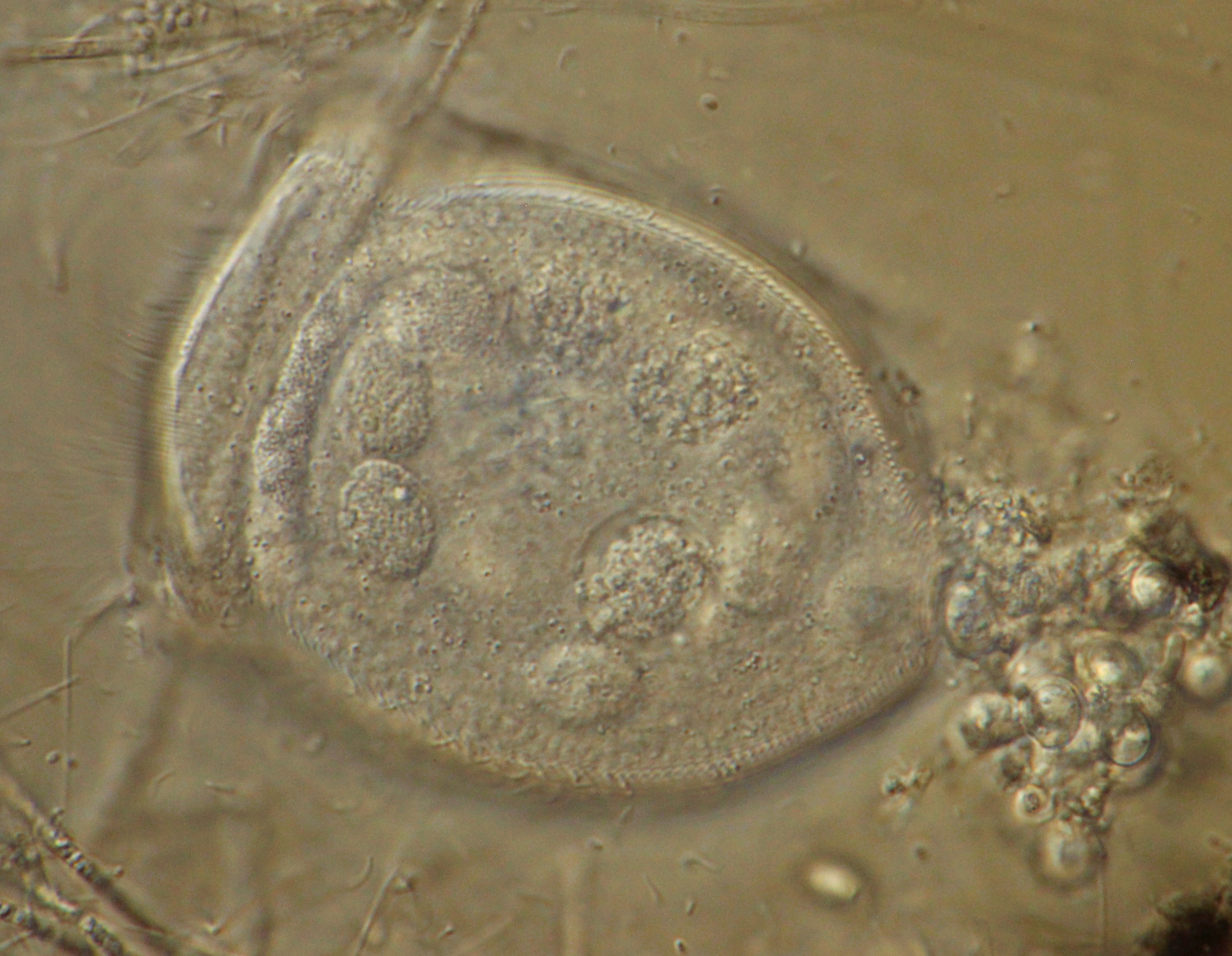
This last image focuses on the food vacuoles. They are the smaller spheres distributed throughout the specimen.
No comments:
Post a Comment